When there is no furcal involvment, these seem to work out OK if the perio heals well.

Online Endodontic Education
When there is no furcal involvment, these seem to work out OK if the perio heals well.
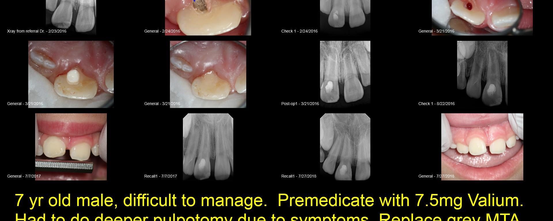
Very young 7 yo male. Original injury Nov 2015. Tooth was restored/bonded and became symptomatic 6 mo later. He saw another endodontist who open the tooth with much difficulty due to patient behavior and uncooperative way. I took over the case, premedicate the pt with valium and eventually removed the old grey MTA because he remained […]
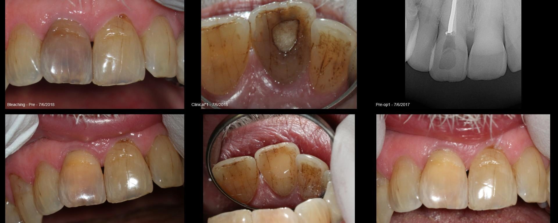
Tough very old discolored tooth. Previous RCT dome over 40 years ago. No pain, completely asymptomatic but just really bad color. Big shot attorney in DC. He was thrilled with the result.
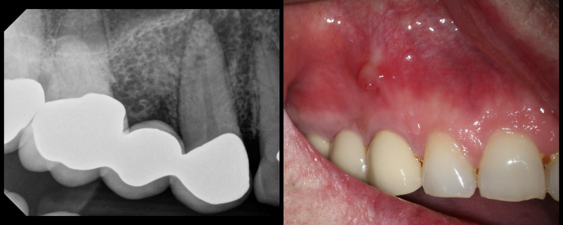
Entire buccal plate missing. CBCT at 5 years suggests most, but not all, bone has regenerated. As canine bridge abutment, it is subjected to some occlusal trauma. gbc

– Sub-gingival caries distal to an isolated #13 – Gingivectomy and isolation done – Caries driven access through the distal (missing #12 and #14 made this approach possible) – No post-endodontic restoration planned as the patient is a partial denture user (maxillary an mandibular) an I don’t think she will generate enough functional load for […]

Thought we would try some practice on JK’s challenge to do a DME on a #2 so I created a deep DO alloy on one of our cadavers and then tried to prep it using his technique. Not perfect, but we are getting better at it. A couple of things: 1. This is way easier […]
#14 RCT Pre-op: Deep sub-gingival distal margin. Gingivectomy followed by margin elevation and isolation. The usual shaping protocol and cleaning protocol. Obturated using WVC. Preserving the dentin bridge that extends buccal to lingual during post-endo prep might be a important consideration in this case.

Clinical Scenario and treatment done DO caries with a deep sub-gingival margin Resorbed apex – Distal root (H/O Orthodontic treatment) Surprisingly had a good tug-back all the way in the distal canals Shaped to 4% 30 Distal and 4% 25 in the mesials Obturation technique – WVC
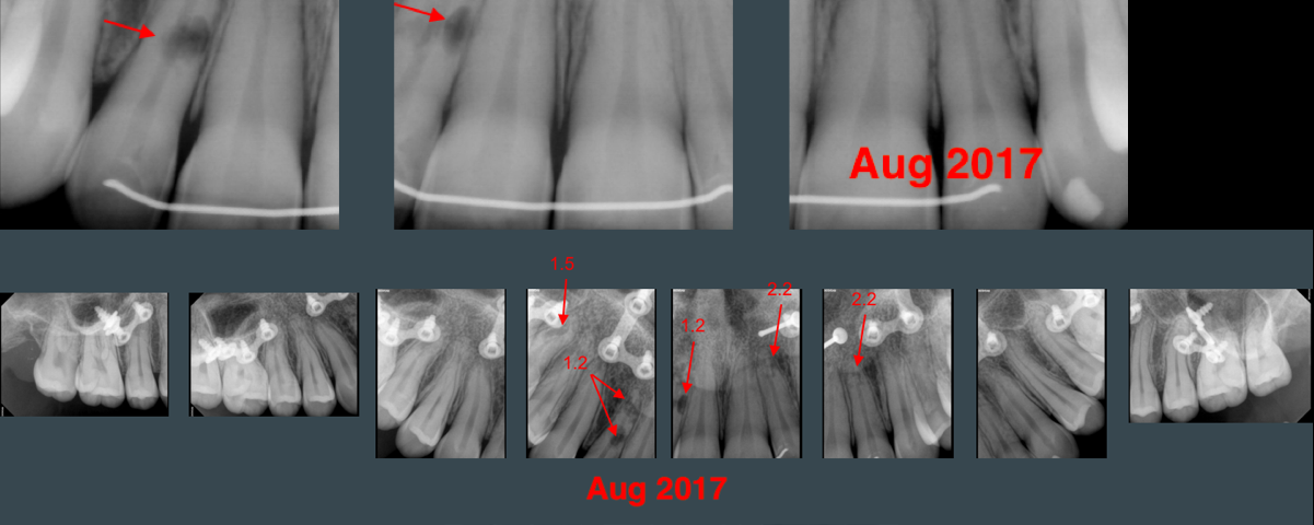
20-yr-old female presents to OMFS w/ CC of open bite in Jan 2013. Orthodontic tx + Orthognathic surgery was planned. Orthodontic tx initiated in Nov 2013. Wisdom teeth extracted in Jan 2016. Orthognathic surgery completed in Aug 2016. I saw the patient on Aug 2017 for assessment of discoloured teeth #1.3 and 2.1 that occurred […]

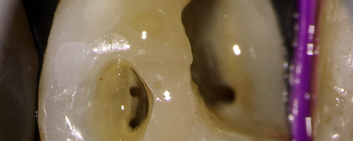
#30 Pulpitis. Caries involving the ML cusp. Caries driven access planned Mb located in the middle of the floor and the dentinal map suggested a ML orifice present almost on the lingual wall of the tooth unlike the usual. I had to stick to what I could see rather than go to the usual co-ordinates to […]